
DOCUMENTED CLINIC CASE
LASIK vision correction
Follow-up one week after surgery
Autorefractor measurements and visual acuity before LASIK and at one week, with preoperative corneal scans. Published with the patient's consent and personal identifiers removed.VISUAL ACUITY
Vision before LASIK and at one week
The following values apply to each eye, as documented by the treating clinician.- Before surgery — without glasses
- 6/60
- Before surgery — with glasses
- 6/6
- At one week — without glasses
- 6/6
BEFORE & AFTER
Autorefractor measurement comparison
Values are transcribed from the summary row of each report. These are autorefractor readings, not visual-acuity measurements or a glasses prescription.| Measurement | Before surgery | At one week |
|---|---|---|
| SphereSPH | −1.00 | +0.25 |
| CylinderCYL | −4.75 | −0.75 |
| AxisAXIS | 166° | 115° |
| Measurement | Before surgery | At one week |
|---|---|---|
| SphereSPH | −1.00 | +0.50 |
| CylinderCYL | −5.25 | −0.50 |
| AxisAXIS | 8° | 129° |
Sphere and cylinder are in dioptres; axis is in degrees.
Original refraction reports
Select an image to open the lighter preview. The full-size original has a separate link with its file size.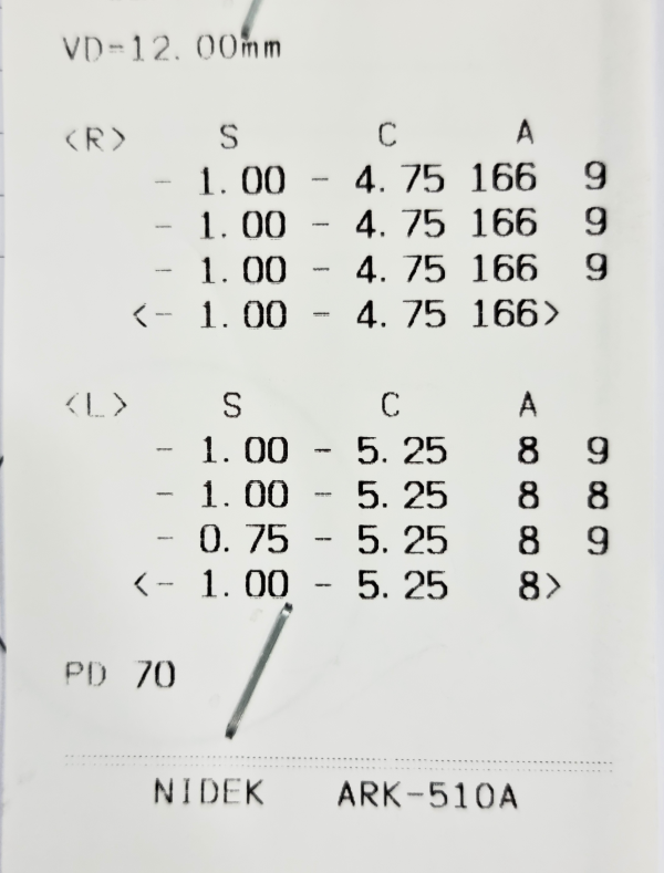 Open preview
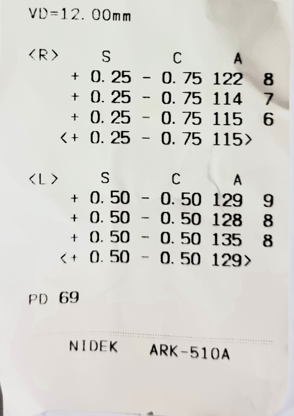 Open preview
PREOPERATIVE EXAMINATIONS
Corneal scans before LASIK
These scans were taken before surgery only. They are not a before-and-after comparison of the cornea. Select an image to open the lighter preview. The full-size original has a separate link with its file size.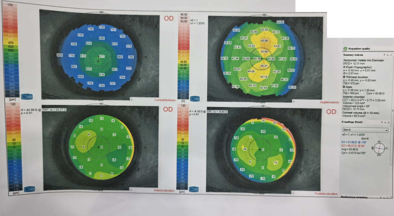 Open preview
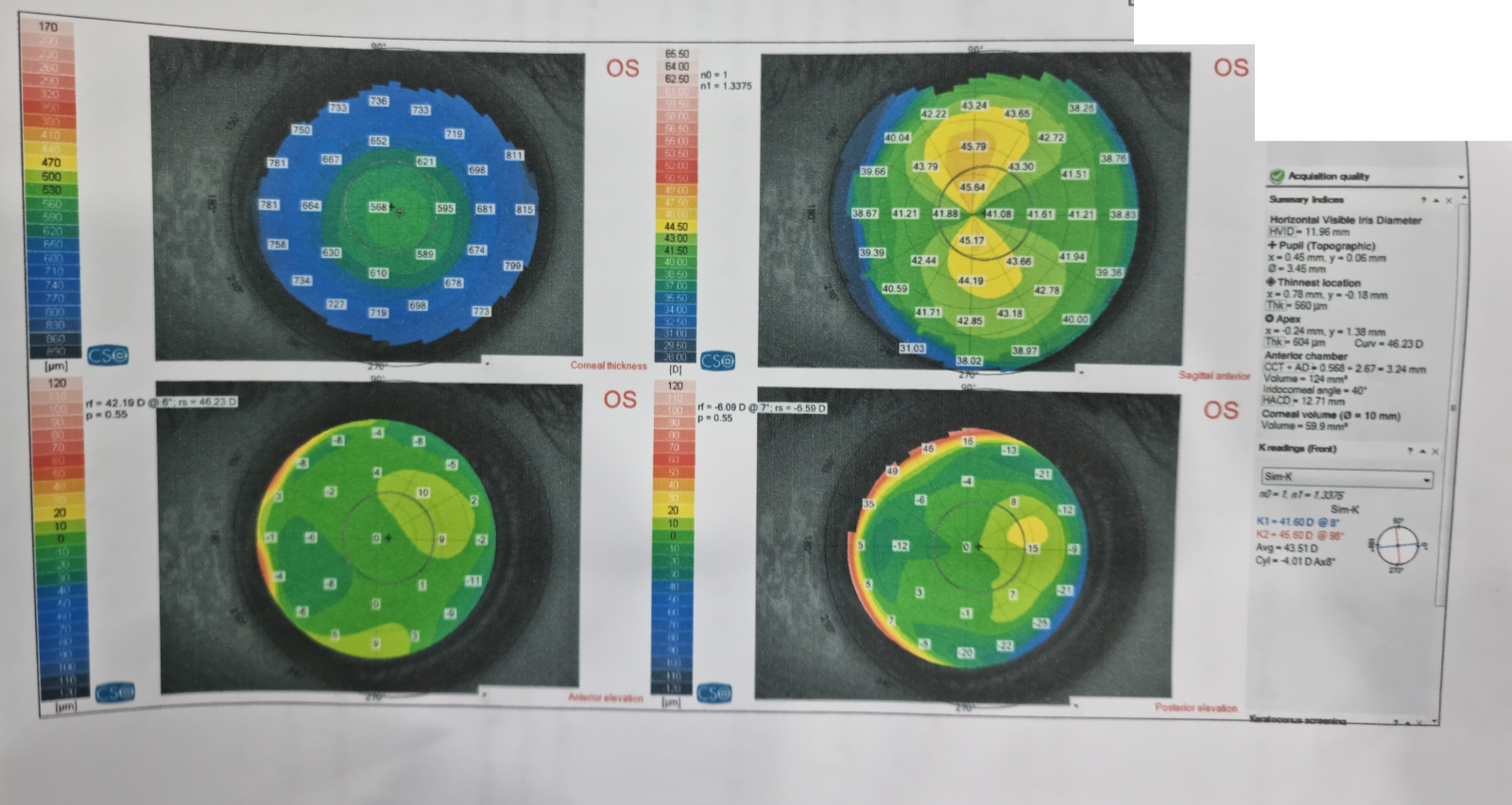 Open preview
PATIENT JOURNEY
Procedure and follow-up details
- Procedure
- LASIK vision correction.
- Displayed follow-up
- One week after surgery.
- Corneal scans
- Preoperative scans of the right and left eyes.
- Patient privacy
- Published with consent and without personal identifiers.